健身人为什么不练腹肌(为什么健身房健身的老练家子最不重视的就是腹肌)
熟悉力量训练/力量举/举重的朋友应该会知道,其实这些体系并不忽视腹肌。相关的文章经常写:“不要忽略腹肌,腹肌在力量训练中非常重要,要收紧核心才能......”。
但这些说法没有足够的科学依据。在全身所有的肌肉中,腹肌和力量水平/力量成绩之间的相关性其实是最低的。
我之前有一篇文章列举了William等人对对20名力量举运动员(其中包含4名世界冠军和3名美国冠军)的研究,发现三大项成绩与他们的肌肉厚度之间呈现极高的正相关性,即肌肉量越大,三大项成绩越高(其他类似的研究说,举起的重量是肌肉量的函数)。
在这篇文章中,成绩与肌肉厚度之间用了一个统计指标,r值。
- r值为0:不相关;
- r值0-0.4:低正相关;
- r值0.4-0.6:中度正相关;
- r值0.6-0.99:强正相关。
在William等人的研究中,这些优秀/顶级力量举运动员的全身所有的肌肉,跟三大项成绩之间都具有非常强的正相关性——但不包括腹肌。
比如第一横行的Forearm(前臂肌肉),与深蹲成绩之间r=0.89,代表前臂肌肉水平和深蹲水平之间,具有极强的正相关性;下面的Biceps(肱二头肌)、Triceps(肱三头肌)等等,都跟三大项成绩之间相关性很高,都在0.7-0.8几左右;Quadriceps(股四头肌)跟卧推之间的正相关性稍微弱一些(r=0.67),可能是因为卧推主要不是靠腿。
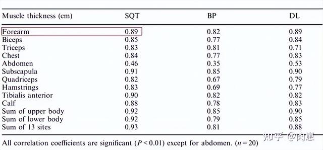
前臂肌肉与三大项成绩之间的强相关性
就连维持平衡的肩袖肌肉—肩胛下肌(Subscapula)与三大项之间的相关性也很高(r=0.85-0.91);与三大项成绩之间相关性最低的是腹肌:abdomen(r=0.35-0.53)。

红框:腹肌与三大项成绩之间的相关性
换句话说,力量举成绩跟全身所有的肌肉体积关联都很大,唯独不包括腹肌。
力量训练根本就不能过分激活腹肌
许多高水平力量运动员都提示大家不要忽视腹肌,包括美国力量举的“教父”路易希蒙。一些美国的力量举文章说他有一种独创的锻炼腹肌的方法:躺在地上,用腹部的力量把放在肚子上的很重的哑铃推开、甚至是抛起来。
但目前的数据并不支持这种观点,力量训练根本不能使劲收缩腹肌。究其原因,一方面因为腹内压和脊柱稳定性的维持主要靠腰带;另一方面因为腹(直)肌的作用主要是脊柱屈,而力量举需要的是脊柱伸[28]。
脊柱的屈和伸,类似于下图描述的概念:
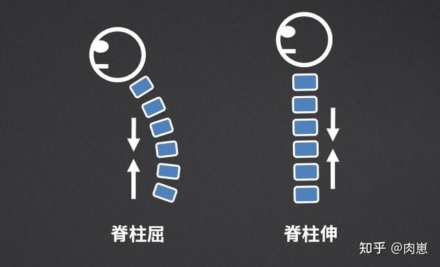
脊柱的屈和伸
在深蹲和硬拉时,人体脊柱可以被视为一个杠杆:重力通过手臂-肩关节,施加在脊柱上,这就造成了一个脊柱屈曲的力、剪切力;如果这个力过大[10],脊柱就会弯曲。

人体杠杆和脊柱
为了避免脊柱弯曲,人体得产生一个让脊柱伸展(挺直)的力。这通过竖脊肌收缩来实现,这就是竖脊肌在解剖上的功能[9,10,30]。
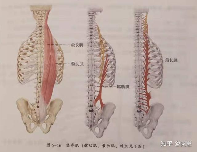
竖脊肌
而腹直肌[31,32,33]发挥脊柱屈曲的作用[34,35],与竖脊肌收缩所产生的力相“对抗”。由于常规的力量训练中深蹲和硬拉时脊柱上承受的力(导致脊柱屈曲的力)已经够大了,人体一方面收缩竖脊肌,一方面也会尽量避免强烈的收缩腹肌。
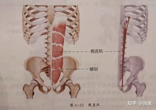
腹直肌
所以常规的力量训练中(特别是深蹲硬拉),竖脊肌的激活必然很高,腹肌激活必然较低。
(肌肉激活度,指的是用相应设备在运动中测得的某个肌肉上的电流。因为肌肉收缩是神经系统放电刺激进行的[1,2,3,4,5,6],测得的电流在基本可以等同于肌肉用力程度[7,8])
这方面的证据比较充分,也比较一致。戴维等人对一群平均体重92kg/身高175cm/前蹲3RM平均125kg的训练者进行测试。

动作示意图
果然,在前蹲和杠铃过顶蹲中,竖脊肌激活很高,腹肌激活很少。


当然,这不是孤证,基本在实践中都会被反复重现。保罗等人研究观察了一群年轻健身者,他们平均21岁,82kg,174cm,具有2年以上(每周至少3次)系统训练经验,测试动作是前蹲和后蹲。
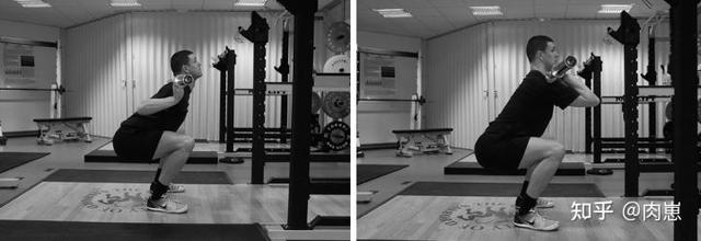
动作示意图
跟之前戴维等的结果类似,前后蹲中腹直肌的激活度很低,约为0.06-0.07左右;竖脊肌的激活水平比腹直肌高得多,约为0.82-1.09。用图形展示较为直观:
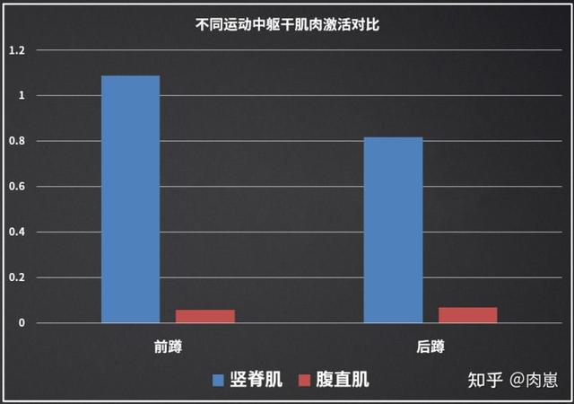
腹肌在前后蹲中的激活都很小
这些数据证明了我们在解剖学功能和运动生物力学层面推测:腹肌在常规力量训练(三大项)中的作用更多是个“拖油瓶”,人体要避免腹肌的过多激活,避免脊柱弯曲。
这也解释了为什么William等人的研究发现,优秀/世界级力量举运动员的腹肌发达程度与三大项之间的关系密切程度最低。
不是说要使劲收紧核心吗?
有些人可能会疑惑,他们看到的文章中说要收紧腹肌,绷紧腰腹。这种说法其实不太准确,主要是相关自媒体的专业度不足造成的。在康复医学领域,腰椎/核心稳定更多是强调竖脊肌/腰部伸肌[11,12]。
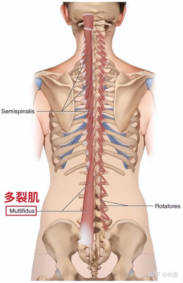
例如多裂肌,大量文献认为它是维持躯干稳定的主要肌肉[13,14,15,16,17,18,19,20,21,22,23,24];针对相应腰部肌肉的训练,被认为是治疗腰疼的重要手段[25,2,627]。
题外话:“卧推最靠肌肉量”不正确
网络和知乎上有一种流行很久的说法:卧推是(三大项中)最靠肌肉量的。
这个说法是错的。
这个说法可能源于是200X年的一篇外国力量举文章,文章说某外国训练者为了达成卧推180kg的挑战(注意,该训练者接受挑战时已经有能力卧推160kg左右),在X个月内并没有狂练卧推(但至少一周练了2次),而是主要靠吃。
大量增加了增加体重和肌肉体积后,该训练者在规定的几个月内提高了20kg卧推,达成了目标。后来这篇文章被翻译到了国内,但是网络上被传的变味了:本来是“卧推很依靠肌肉量”,结果成了“卧推是三大项中最依赖肌肉量的”。
卧推靠不靠肌肉量呢?毫无疑问,肯定的。在William等人的数据中,这关系就明摆着。
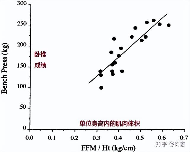
但是卧推并不是与肌肉量关系最大的项目。
在William等人的数据中,最后一行是“13个部位的肌肉总和”。我们清楚地看到,全身肌肉量跟深蹲之间的相关性最强(r=0.93),卧推(r=0.81)和硬拉(r=0.88)分别是排在后面。
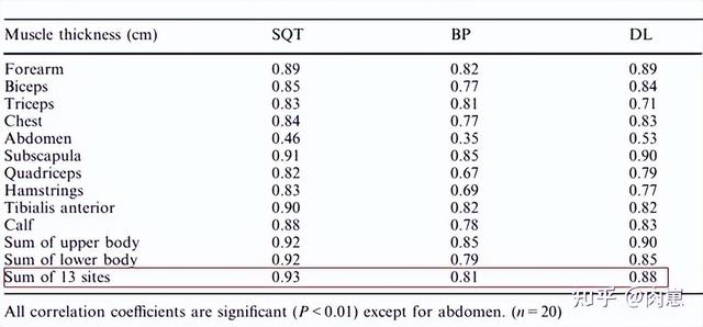
这些数据表明,深蹲才是最吃肌肉量的项目。虽然科学一般不说个例,但我自己和周围的朋友的个人经验跟这个数据是吻合的。当因为减脂、停练、工作等各种原因体重明显下降时,成绩掉得最多的是深蹲,卧推的下降想对没那么多。
总之
1. 力量训练中,脊柱主要是伸,竖脊肌激活很高,腹肌不会被大幅度激活,强大的力量很可能不需要太强的腹肌
2. 力量举三大项中最吃肌肉量的项目,是深蹲,而不是卧推
3. “收紧核心”在腰椎稳定中更多指的是收紧竖脊肌/背部-腰部肌肉
References
1. Huxley AF, Niedergerke R (1954) Structural changes in muscle during contraction. Interference microscopy of living muscle fibres. Nature 173:971–973.
2. Huxley HE, Hanson J (1954) Changes in cross-striations of muscle during contraction and stretch and their structural implications. Nature 173:973–976.
3. Huxley HE (1969) The mechanism of muscular contraction. Science 164:1356–1366.
4. Huxley AF, Simmons RM (1971) Proposed mechanism of force generation in striated muscle.
5. Rayment I, Holden HM, Whittaker M, Yohn CB, Lorenz M, Holmes KC, Milligan RA (1993) Structure of the actin-myosin complex and its implications for muscle contraction. Science 261:58–65.
6. Rayment I, Rypniewski WR, Schmidt-B?se K, Smith R, Tomchick DR, Benning MM, Winkelmann DA, Wesenberg G, Holden HM (1993) Three-dimensional structure of myosin subfragment-1: a molecular motor. Science 261:50–58.
7. Nicolas Babault 1 , Michel Pousson, Anne Michaut, Yves Ballay, Jacques Van Hoecke.EMG activity and voluntary activation during knee-extensor concentric torque generation.Eur J Appl Physiol. 2002 Apr;86(6):541-7.
8. F Bellemare, J J Woods, R Johansson, B Bigland-Ritchie.Motor-unit discharge rates in maximal voluntary contractions of three human muscles.J Neurophysiol. 1983 Dec;50(6):1380-92.
9. S L Delp 1 , S Suryanarayanan, W M Murray, J Uhlir, R J Triolo.Architecture of the rectus abdominis, quadratus lumborum, and erector spinae.J Biomech. 2001 Mar;34(3):371-5.
10. Christopher J Colloca 1 , Richard N Hinrichs.The biomechanical and clinical significance of the lumbar erector spinae flexion-relaxation phenomenon: a review of literature.J Manipulative Physiol Ther. 2005 Oct;28(8):623-31.
11. Karen P Barr 1 , Miriam Griggs, Todd Cadby.Lumbar stabilization: core concepts and current literature, Part 1.Am J Phys Med Rehabil. 2005 Jun;84(6):473-80.
12. Karen P Barr 1 , Miriam Griggs, Todd Cadby.Lumbar stabilization: a review of core concepts and current literature, part 2.Am J Phys Med Rehabil. 2007 Jan;86(1):72-80.
13. Hides J, Stokes MJ, Saide M, Jull GA, Cooper DH. Evidence of lumbar multifidus muscle wasting ipsilateral to symptoms in patients with acute/subacute low back pain. Spine. 1994;19(2):165–172.
14. Hodges PW, Holm AK, Hansson T, Holm S. Rapid atrophy of the lumbar multifidus follows experimental disc or nerve root injury. Spine. 2006;31(25):2926–2933.
15. Hodges PW, Danneels L. Changes in structure and function of the back muscles in low back pain: different time points, observations, and mechanisms. J Orthop Sports Phys Ther. 2019;49(6):464–476.
16. Wei-ye Chen, Kuan Wang, Wei-an Yuan, Hong-sheng Zhan.Relationship between lumbosacral multifidus muscle and lumbar disc herniation.Zhongguo Gu Shang. 2016 Jun;29(6):581-4.
17. Hicks GE, Fritz JM, Delitto A, McGill SM. Preliminary development of a clinical prediction rule for determining which patients with low back pain will respond to a stabilization exercise program. Arch Phys Med Rehabil. 2005;86(9):1753–1762.
18. Solomonow M, Zhou BH, Harris M, Lu Y, Baratta RV. The ligamento-muscular stabilizing system of the spine. Spine. 1998;23(23):2552–2562.
19. Wilke HJ, Wolf S, Claes LE, Arand M, Wiesend A. Stability increase of the lumbar spine with different muscle groups. A biomechanical in vitro study. Spine. 1995;20(2):192–198.
20. Wagner H, Anders C, Puta C, Petrovitch A, Mörl F, Schilling N, et al. Musculoskeletal support of lumbar spine stability. Pathophysiology. 2005;12(4):257–265.
21. Hay EM, Dunn KM, Hill JC, Lewis M, Mason EE, Konstantinou K, et al. A randomised clinical trial of subgrouping and targeted treatment for low back pain compared with best current care. The STarT Back Trial Study Protocol. BMC Musculoskelet Disord. 2008;9:58.
22. Hill JC, Dunn KM, Main CJ, Hay EM. Subgrouping low back pain: a comparison of the STarT Back Tool with the Orebro Musculoskeletal Pain Screening Questionnaire. Eur J Pain. 2010;14(1):83–89.
23. Hill JC, Whitehurst DG, Lewis M, Bryan S, Dunn KM, Foster NE, et al. Comparison of stratified primary care management for low back pain with current best practice (STarT Back): a randomised controlled trial. Lancet. 2011;378(9802):1560–1571.
24. Stanton TR, Hancock MJ, Apeldoorn AT, Wand BM, Fritz JM. What characterizes people who have an unclear classification using a treatment-based classification algorithm for low back pain? A cross-sectional study. Phys Ther. 2013;93(3):345–355.
25. Ko KJ, Ha GC, Yook YS, Kang SJ. Effects of 12-week lumbar stabilization exercise and sling exercise on lumbosacral region angle, lumbar muscle strength, and pain scale of patients with chronic low back pain. J Phys Ther Sci. 2018;30(1):18–22.
26. Bagheri R, Takamjani IE, Dadgoo M, Sarrafzadeh J, Ahmadi A, Pourahmadi MR, et al. A protocol for clinical trial study of the effect of core stabilization exercises on spine kinematics during gait with and without load in patients with non-specific chronic low back pain. Chiropr Man Ther. 2017;25:31.
27. Sung PS, Yoon B, Lee DC. Lumbar spine stability for subjects with and without low back pain during one-leg standing test. Spine. 2010;35(16):E753–E760.
28. H Kalimo, J Rantanen, T Viljanen, S Einola.Lumbar muscles: structure and function.Ann Med. 1989 Oct;21(5):353-9.
29. N Bogduk.A reappraisal of the anatomy of the human lumbar erector spinae.J Anat. 1980 Oct;131(Pt 3):525-40.
30. Thomas M Greiner 1 , M Elizabeth Bedford, Robert A Walker.Variability in the human m. spinalis capitis and cervicis: frequencies and definitions.Ann Anat. 2004 Apr;186(2):185-91.
31. Fabien Fredon, Jérémy Hardy, Mélanie Germain, Emma Vincent-Viry, Abdelkader Taïbi, Jacques Monteil, Christian Mabit, Denis Valleix, Sylvaine Durand-Fontanier.Correlations of the rectus abdominis muscle anatomy with anthropometric measurements.Surg Radiol Anat. 2021 Apr;43(4):589-593.
32. Justin M Broyles, Mark D Schuenke, Sima R Patel, Caroline M Vail, Heather V Broyles, A Lee Dellon.Defining the Anatomy of the Tendinous Intersections of the Rectus Abdominis Muscle and Their Clinical Implications in Functional Muscle Neurotization.Ann Plast Surg. 2018 Jan;80(1):50-53.
33. Denise de Almeida Mendes 1 , Fábio Xerfan Nahas, Daniela Francescato Veiga, Fernando Vilela Mendes, Ricardo Góes Figueiras, Heitor Carvalho Gomes, Pedro Bins Ely, Neil Ferreira Novo, Lydia Masako Ferreira.Ultrasonography for measuring rectus abdominis muscles diastasis.cta Cir Bras. May-Jun 2007;22(3):182-6.
34. Flint, M. M. Abdominal muscle involvement during performance of various forms of sit-up exercises: electromyographic study.Am. J. Phys. Med. 44:224-234, 1965.
35. Walters, C. E. and M. J. Partridge. Electromyographic study of the differential action of the abdominal muscles during exercise.Am. J. Phys. Med. 36:259-268, 1957.
,


- 内双眼皮怎么快速变外双(不是说通过双眼皮就可以改善的)
- win7最近老是未响应(win7闪屏后未响应解决步骤)
- 怎么在手机浏览器上登录qq(怎样跳过短信验证)
- excel一个表格内求和(2022年4种最全求和方法)
- 埃迪斯科文大学有什么优势(埃迪斯科文大学为什么这么好)
- 酸辣鲫鱼好吃又下饭(酸辣爽口又营养美味)
- 冥想的好处在哪里(经常练习冥想对我们有哪些好处)
- 笔记本电脑wifi不能启动怎么办(专业IT运维服务商教你自动连接)
- 哔哩哔哩为啥有的番剧不能承包(承包番剧或开通大会员免广告)
- 三色藜麦的功效与作用及食用方法(三色藜麦饭团营养又健康)
- 轮虫对水体有什么的作用(它有很多好处)
- 一个互联网运营需要的技能(合格的运营人必备)
- 24季节气排序(24节气时间日期对照表查询)
- 蒸的苹果是怎么做的(想不到作用这么大)
- 离婚起诉状女方怎么写简单有效(新婚姻法女方怎样写起诉离婚书)
- 小米相机ai模式是什么意思(小米最强的功能)
- 抖音画面不流畅怎么解决(你会用吗)
- 考试做题的方法和技巧(保持年级前3名)